Accreditation
CVT Vascular Lab is accredited by the Intersocietal Accreditation Commission (IAC). IAC offers accreditation for each of the testing areas that the vascular laboratory performs. CVT Vascular Lab first sought and obtained IAC accreditation in 1993 and has continually maintained accreditation since that time.
IAC accreditation is recognized as certification that the Vascular Laboratory meets established standards of quality in vascular testing. The accreditation process considers what kind of equipment a laboratory uses, how it is maintained, testing protocols, reporting procedures, training and qualification of the staff, and the quality-assurance processes that are in place. Meeting or exceeding the nationally recognized standards of quality, vascular testing at CVT Vascular Lab is done by trained and credentialed vascular technologists using state-of-the-art ultrasound imaging and noninvasive vascular testing systems.
Accreditation is granted only to those facilities that are found to be providing quality patient care, in compliance with the IAC Standards. Once granted, IAC accreditation is valid for a period of three years, after which time the facility must undergo a repeat evaluation.
Lab Technology
Ultrasound duplex scanning is the workhorse technology for the Vascular Laboratory. Duplex scanning is non-invasive, safe, and cost-effective. It can be used for vascular disease screening, but it can also provide a definitive diagnosis in many cases.
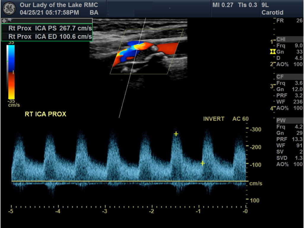
Duplex scanning uses two-dimensional, grayscale, B-mode imaging to create a picture of vessel anatomy.
Imaging can also characterize plaque, thrombus or other vessel pathology. Doppler ultrasound evaluation of blood-flow velocities is the other component of duplex scanning. Flow velocity information can be read from a Doppler waveform or displayed as a color overlay on the grayscale image.
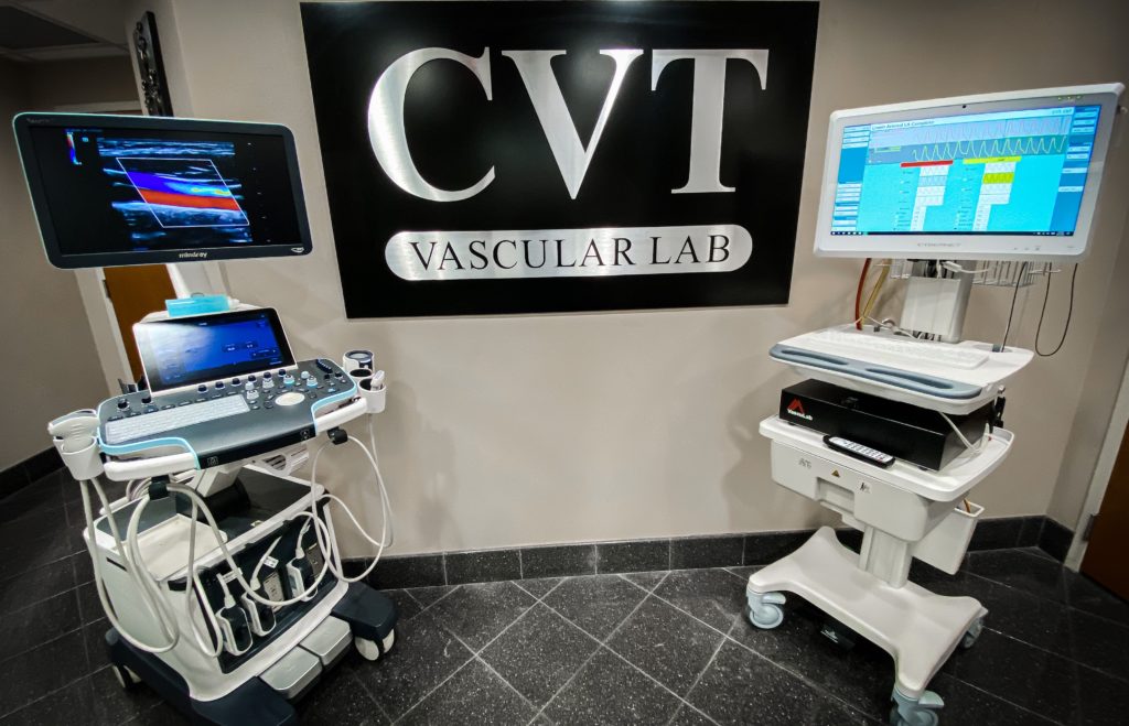
CVT Vascular Lab uses a suite of full-featured ultrasound systems with state-of-the-art imaging technologies.
These ultrasound systems are used for many types of studies. Electronic image recording and storage on our PACS system allows clinicians convenient and secure access to vascular studies from any network workstation.
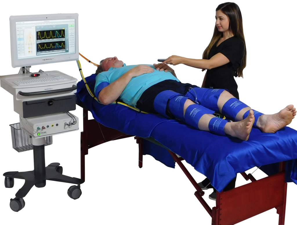
Testing for upper- and lower-extremity arterial disease commonly includes the use of physiologic testing methods. These tests include limb or digit pressure measurements with pneumatic cuffs and Doppler flow detectors, plethysmography (measurements of volume changes due to pulsatile blood flow), and other techniques. These tests can also be performed before and after treadmill exercise, which may be helpful in the evaluation of patients with pain while walking.